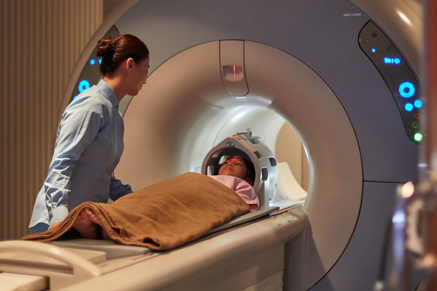
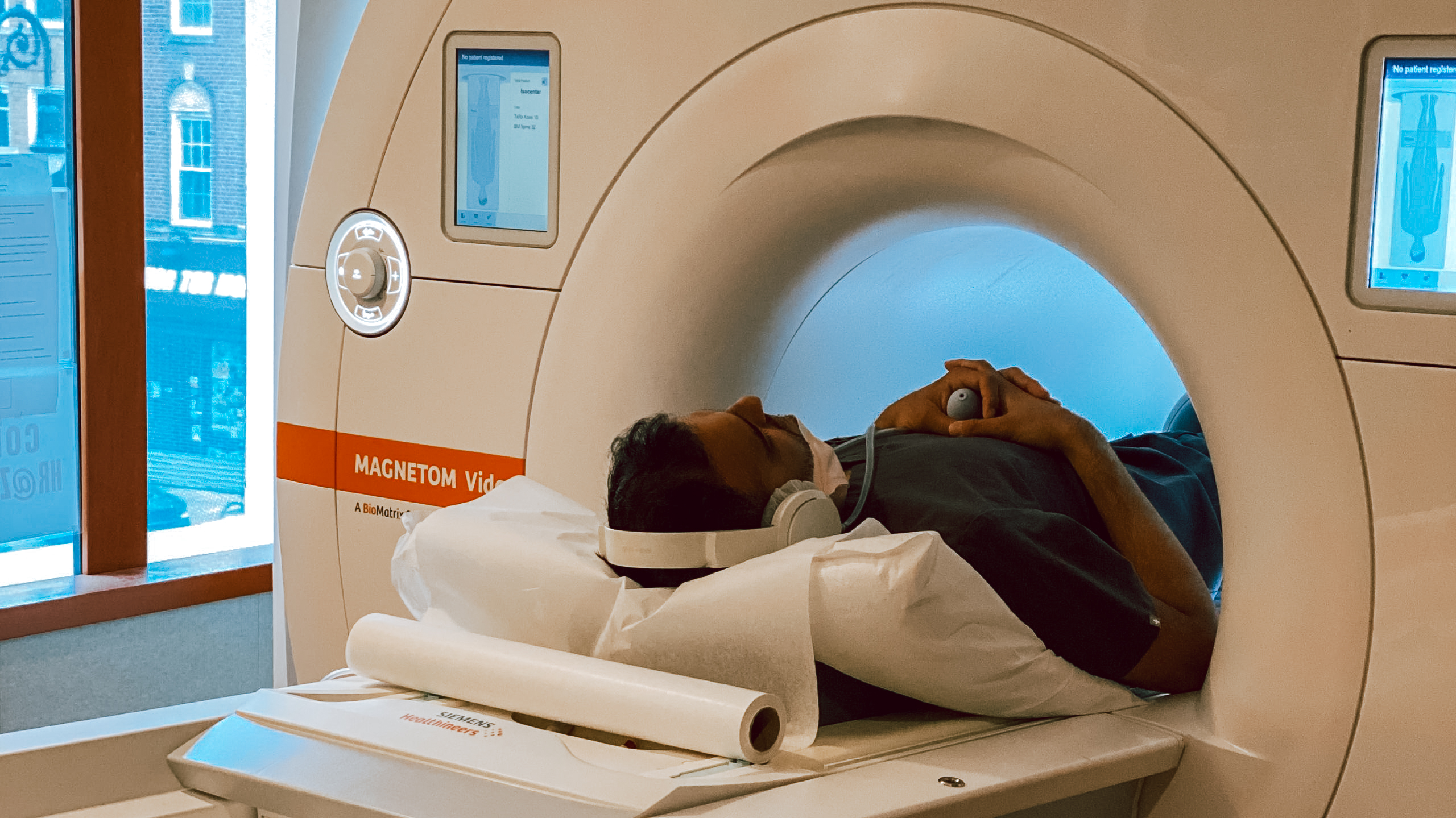
Magnetic Resonance Imaging (MRI) has revolutionized the field of medical imaging, providing detailed and non-invasive visualization of internal structures.
This article explores the history, mechanics, and various applications of MRI.
Furthermore Lead glass supplier, it delves into the benefits and potential risks associated with this imaging technique.
By using a technical and analytical approach, this concise introduction aims to provide a comprehensive overview of MRI for an audience seeking accurate information and a deeper understanding of this vital diagnostic tool.

History of MRI
The history of MRI dates back to the 1970s when it was first developed as a medical imaging technique. Magnetic Resonance Imaging (MRI) has seen a remarkable evolution over the years, revolutionizing medical diagnosis.
Initially, MRI scanners were large and required extensive cooling systems. However, advancements in technology have resulted in the development of smaller, more efficient MRI machines accentrix company limited. These machines utilize a strong magnetic field and radio waves to generate detailed images of the body’s internal structures.
The introduction of advanced imaging sequences, such as T1-weighted, T2-weighted, and diffusion-weighted imaging, has further enhanced the diagnostic capabilities of MRI. Furthermore, the incorporation of contrast agents has allowed for improved visualization of blood vessels and abnormalities.
With its non-invasive nature and ability to produce high-resolution images, MRI has become an indispensable tool in the field of medical imaging, facilitating accurate diagnoses and guiding effective treatments.
How MRI Works
MRI machines generate detailed images of the body by using a strong magnetic field and radio waves. This non-invasive imaging technique has revolutionized medical diagnostics by providing high-resolution images of soft tissues, organs, and even the brain.
Recent advancements in MRI technology have improved image quality, reduced scan times, and expanded the range of applications. For example, the development of stronger magnets and improved gradient systems has led to higher signal-to-noise ratios and more detailed images. Additionally, the incorporation of advanced software algorithms and machine learning techniques has enhanced image reconstruction and analysis.
In the future, further developments in MRI are expected, such as the integration of artificial intelligence for automated image interpretation and the development of portable MRI scanners that can be used in various clinical settings. These advancements will continue to enhance patient care and contribute to the field of medical imaging.
Applications of MRI
Applications of magnetic resonance imaging encompass a wide range of medical specialties, including neurology, orthopedics, cardiology, and oncology.
MRI is a powerful diagnostic tool that provides detailed images of the body’s internal structures and functions.
In neurology, MRI is used to detect and monitor brain tumors, multiple sclerosis, and other neurological disorders.
In orthopedics, MRI helps diagnose and assess injuries to bones, joints, and soft tissues, such as ligament tears and cartilage damage.
Cardiologists use MRI to evaluate heart function, detect abnormalities, and assess the extent of cardiac damage.
In oncology, MRI plays a crucial role in detecting and staging tumors, guiding biopsies, and monitoring treatment response.
Advancements in MRI technology, such as higher field strengths and improved image quality, have greatly enhanced its diagnostic capabilities.
Future developments in MRI research aim to further improve image resolution, reduce scan times, and enhance the integration of functional imaging techniques, ultimately leading to more precise and personalized patient care.
Benefits of MRI

MRI offers numerous advantages in the field of medical imaging, providing non-invasive and highly accurate diagnostic information that aids in the detection and management of a wide range of medical conditions. Advancements in MRI technology have led to improved image quality and faster scan times, allowing for more efficient and precise diagnoses.
The development of high-field MRI systems has further enhanced the capabilities of this imaging modality by increasing spatial resolution and signal-to-noise ratio. Additionally, the incorporation of advanced imaging techniques such as functional MRI (fMRI) and diffusion-weighted imaging (DWI) has provided valuable insights into brain function and tissue microstructure.
Future developments in MRI aim to overcome current limitations, such as motion artifacts and the need for contrast agents, by exploring novel imaging sequences and hardware innovations. With ongoing research and technological advancements, MRI will continue to play a crucial role in the diagnosis and management of various medical conditions.
Potential Risks of MRI
While MRI is generally considered safe, there are potential risks associated with the use of strong magnetic fields, such as the heating of implanted metal devices or the presence of ferromagnetic objects in the patient’s body. These risks highlight the importance of MRI safety protocols and thorough patient screening before undergoing an MRI examination.
One potential risk involves the heating of implanted medical devices, such as pacemakers or cochlear implants, due to the interaction with the strong magnetic field. This can lead to tissue damage or malfunction of the device.
Another risk is the presence of ferromagnetic objects in the patient’s body, such as metal fragments or tattoos containing metallic pigments. These objects can be attracted to the magnetic field, causing movement or displacement within the body, potentially resulting in injury.
It is crucial for radiologists and technologists to be aware of these risks and take appropriate measures to ensure patient safety during MRI procedures. Additionally, the use of MRI contrast agents, which are substances injected into the patient’s body to enhance the visibility of certain tissues, can also present risks, such as allergic reactions or nephrogenic systemic fibrosis in patients with impaired kidney function. Close monitoring and proper administration of contrast agents are essential to minimize these risks.
Overall, while MRI is a valuable diagnostic tool, it is important to be aware of and manage the potential risks associated with its use.
Conclusion
In conclusion, MRI has revolutionized the field of medical imaging by providing detailed and accurate images of the body’s internal structures. Its non-invasive nature and ability to capture images in different planes make it a valuable tool in diagnosing various conditions.
While MRI offers numerous benefits, such as improved visualization and early detection of abnormalities, it is important to consider potential risks, such as the use of contrast agents and the presence of magnetic fields.
Overall, MRI continues to play a crucial role in medical diagnostics and research.
